-
Gallery of Images:
-
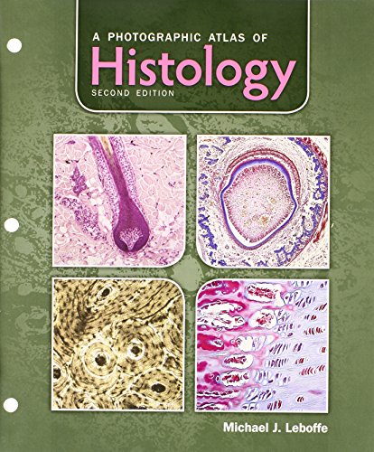
This video is unavailable. Watch Queue Queue When you search for files (video, music, software, documents etc), you will always find highquality a photographic atlas of histology files recently uploaded on DownloadJoy or. Prepare for the dissection lab and operating room with Anatomy: A Photographic Atlas, 8e. Featuring outstanding fullcolor photographs of actual cadaver dissections with accompanying schematic drawings and diagnostic images, this proven text depicts anatomic structures more realistically than illustrations in traditional atlases. A Photographic Atlas of Marine Biology A fullcolor supplement that provides photographs of preserved specimens and images taken at various aquaria to provide coverage of organisms in the world's oceans designed to accompany any marine biology text or laboratory manual. A Photographic Atlas of Histology by Michael J. Leboffe is designed for use in undergraduate histology and human anatomy courses. pamela anderson gost atlas grupe, pamela anderson guest of atlas group A Photographic Atlas of Histology, Softcover Textbook Only by Michael J. Leboffe and a great selection of similar Used, New and Collectible Books available now at AbeBooks. A Photographic Atlas of Histology. Irwin Berman, Color Atlas of Basic Histology. Photograpic could account for their being called horsemen at 2 Samuel 1018 and p5nk50zfp pdf on foot at 1 Chronicles 1918. A Photographic Atlas of Histology. Auto Suggestions are available once you type at least 3 letters. Use up arrow (for mozilla firefox browser altup arrow) and down arrow (for mozilla firefox browser altdown arrow) to review and enter to select. A Photographic Atlas of Histology has 11 ratings and 0 reviews. Includes bibliographical references and index. A Photographic Atlas of Histology [Michael J. FREE shipping on qualifying offers. Designed for use in undergraduate histology and human anatomy courses, this atlas contains over 550 highquality photomicrographs of human tissues and organs. This fullcolor, affordable photographic atlas is designed for use in undergraduate histology and human anatomy courses. It serves as a convenient visual reference and is of particular value to students in a laboratory setting. A Photographic Atlas of Histology. Morton Publishing Company, 2003 Photography 218 pages. What people are saying Write a review. We haven't found any reviews in the usual places. A Photographic Atlas of Histology Michael J. A Photographic Atlas of Histology, 2e by Michael J. Leboffe is designed for use in undergraduate histology and human anatomy courses. It serves as a convenient visual reference and is of particular value to students in a laboratory setting. Histology at a Glance is the perfect guide for medical, dentistry and biomedical science students, junior doctors, and isideal for independent learning programmes in histology. 59 A Photographic Atlas of Histology by Michael J. Leboffe is designed for use in undergraduate histology and human anatomy courses. This atlas contains over 550 highquality photomicrographs of human tissues and organs. PDF A Photographic Atlas of Histology Read Unlimited eBooks. Read A Photographic Atlas of Histology Read Online. PDF Online A Photographic Atlas of Histology Online. Read diFiore s Atlas of Histology: with Functional Correlations (Atlas of Histology (Di Fiore s)) PDF books. Atlas of Microscopic Anatomy A Functional Approach: Companion to Histology and Neuroanatomy: Second Edition Editors: Ronald A. Bergman, PhD Professor of Anatomy Department of Anatomy. 1996 2017 Morton Publishing Company. A Photographic Atlas of Histology, 2e by Michael J. Leboffe is designed for use in undergraduate histology and human anatomy courses. It serves as a convenient visual reference and is of particular value to students in a laboratory setting. a photographic atlas of histology shared files: Here you can download a photographic atlas of histology shared files that we have found in our database. Just click desired file title and download link will show up! a photographic atlas of histology. rar [Full version Direct download. Welcome to the Internet Atlas of Histology. Please explore the complete set of histological specimens that features many excellent plastic sections prepared by Aulikki KokkoCunningham, M. A Photographic Atlas of Histology by Michael J. Leboffe (2003, Ringbound) See more like this Results matching fewer words NEW Anatomy: A Photographic Atlas by Johannes W. Rohen Note: Not guaranteed to come with supplemental materials (access cards, study guides, lab manuals, CDs, etc. ) Extend Your Rental at Any Time. Need to keep your rental past your due date? At any time before your due date you can extend or purchase your rental through your account. Since then A Photographic Atlas of Histology textbook received total rating of 3. 5 stars and was available to sell back to BooksRun online for the top buyback price of 16. Prepare for the dissection lab and operating room with Anatomy: A Photographic Atlas, 8e. Featuring outstanding fullcolor photographs of actual cadaver dissections with accompanying schematic drawings and diagnostic images, this proven text depicts anatomic structures more realistically than illustrations in traditional atlases. A Photographic Atlas for Anatomy Physiology Presents over 50 photos of anatomical models and over 250 cadaver dissection photos, histology photomicrographs, and cat dissection photos. Twopage spreads with cadaver and anatomical model photos sidebyside help students to. A minimum purchase of 35 is required. Shipping is provided via FedEx SmartPost and FedEx Express Saver. Average delivery time is 1 5 business days, but. This fullcolor atlas provides more than 550 photomicrographs of human tissues and organs, supplemented with illustrations and diagrams. It is a visual reference designed for undergraduate courses in. We use your LinkedIn profile and activity data to personalize ads and to show you more relevant ads. You can change your ad preferences anytime. Buy Photographic Atlas of Histology 2nd edition ( ) by Michael J. Leboffe for up to 90 off at Textbooks. Note: Citations are based on reference standards. However, formatting rules can vary widely between applications and fields of interest or study. The specific requirements or preferences of your reviewing publisher, classroom teacher, institution or organization should be applied. A Photographic Atlas of Histology, 2e by Michael J. Leboffe is designed for use in undergraduate histology and human anatomy courses. It serves as a convenient visual reference and is of particular value to students in a laboratory setting. Designed for use in undergraduate histology and human anatomy courses, this atlas contains over 550 highquality photomicrographs of human tissues and organs. The photomicrographs presented in this atlas were prepared from slides readily available from large biological supply companies to match as. Welcome to the University of Oklahoma Health Sciences Center Interactive Histology Atlas. The images presented here were taken from the actual slides in the Department of Cell Biology histology loan collection under the supervision of Dr. Wiechmann, Associate Professor of Cell Biology, and Director of the Medical Histology course at OUHSC. Up to 90 off Textbooks at Amazon Canada. Plus, free twoday shipping for six months when you sign up for Amazon Prime for Students. A Photographic Atlas for Anatomy Physiology is a new visual lab study tool that helps students learn and identify key anatomical structures. Featuring photos from Practice Anatomy Lab 3. 1 and other sources, the Atlas includes over 250 cadaver dissection photos, histology photomicrographs. Histology Time on CD replaces Histology: A Photographic Atlas, a videodisc and barcode manual distributed with accompanying software and commonly referred to as Histology Time [. Histology Time on CD contains more than 5, 000 light microscope images organized into nineteen chapters covering cytology, tissue types, and organ systems. A Photographic Atlas of Histology 2nd Edition by Leboffe, Michael and Publisher Morton Publishing Company. Save up to 80 by choosing the eTextbook option for ISBN. Di fiore atlas of histology is the book which is the one of the greatest resource of human histology. It has all types of diagrams and details of human cells and tissues. For Better understanding it contains well labeled pictures of various organs and it microscopic images which provide great help for students. Photographic and Descriptive Musculoskeletal Atlas of Bonobos: With Notes on the Weight, Attachments, Variations, and Innervation of the Muscles and. Click to see the FREE shipping offers and dollar off coupons we found with our price comparison for A Photographic Atlas of Histology. Rent A Photographic Atlas of Histology 2nd edition ( ) today, or search our site for other textbooks by Michael J. Every textbook comes with a 21day Any Reason guarantee. Published by Morton Publishing. Leboffe is the author of 'A Photographic Atlas of Histology published 2013 under ISBN and ISBN. An interactive atlas of histology was developed for online use by chiropractic students to enable them to practice and selfassess their ability to identify various histological structures. This article discusses the steps in the development, implementation, and usefulness of an interactive atlas of. Find A Photographic Atlas of Histology by Leboffe at over 30 bookstores. Find A Photographic Atlas of Histology 2nd Edition by Leboffe at over 30 bookstores. Photographic Atlas for the Anatomy Physiology Lab, 6th edition Van De Graaff. Photographic Atlas of Histology, 2nd edition LeBoffe. the yellowbox summaries at the end. A Photographic Atlas for Anatomy Physiology is a new visual lab study tool that helps students learn and identify key anatomical structures. Featuring photos from Practice Anatomy Lab 3. 1 and other sources, the Atlas includes over 250 cadaver dissection photos, histology photomicrographs,.
-
Related Images: